
Tattoo Ink Concerns in Laser Tattoo Removal
 Faramarz Rafie MD / DBA – Vancoderm Academy and College [VDA] – Vancoderm Clinic [VDCMed]
Impurities, Mixed Colors, and Clinical Challenges
Laser tattoo removal is widely regarded as the gold standard for removing unwanted tattoos. However, treatment outcomes are not determined by laser technology alone. Tattoo ink composition—including impurities, mixed pigments, and undocumented ingredients—plays a critical role in safety, efficacy, and predictability of results. Understanding these factors is essential for both practitioners and patients.
Tattoo Ink Composition:
Tattoo ink is not a single, standardized substance. Once deposited into the dermis, it becomes a permanent intradermal foreign material composed of pigments, carriers, and secondary contaminants. From a medical and laser-tissue interaction perspective, the chemical nature, particle size, and stability of these components directly influence inflammatory response, laser absorption, and treatment outcomes.
Pigments: Organic vs Inorganic
Tattoo pigments are responsible for color and are broadly classified into organic and inorganic compounds.
Inorganic Pigments
Traditionally used and still common in black, red, yellow, and flesh-tone inks:
- Carbon black (soot-based): highly stable, strong laser absorber
- Iron oxides (Fe₂O₃, Fe₃O₄): red, brown, cosmetic tattoos
- Titanium dioxide (TiO₂): white and pastel shades
- Chromium oxide: green
- Cobalt salts: blue
Clinical relevance:
- Inorganic pigments are generally more thermally stable
- Iron oxide and titanium dioxide may undergo reduction reactions under laser exposure, causing paradoxical darkening
- Heavy metal content increases risk of delayed hypersensitivity and granulomatous reactions
Organic Pigments
Modern tattoos frequently use synthetic organic pigments, especially azo dyes and polycyclic compounds:
- Azo pigments (reds, yellows, oranges)
- Phthalocyanines (blues and greens)
- Quinacridones (violet, magenta)
Clinical relevance:
- Often produce smaller particle fragments during laser treatment
- May degrade into aromatic amines, some of which are potentially toxic or carcinogenic
- Exhibit variable wavelength absorption, complicating laser selection
Particle Size and Dermal Distribution of Tattoo Pigments
The behavior of tattoo ink during laser removal is fundamentally governed by particle size, aggregation state, and spatial distribution within the dermis. These parameters directly influence laser–tissue interaction, immune clearance, and the risk of adverse effects. Tattoo pigment does not exist as free-floating material but as a dynamic intradermal foreign-body system integrated into dermal structures.
1. Particle Size Spectrum and Physical Characteristics
Tattoo pigments demonstrate a polydisperse size distribution, typically ranging from:
-
Nanoparticles: ~20–100 nm
-
Submicron particles: 100–1000 nm
-
Microparticles: 1–5 µm or larger
Key medical implications:
-
Nanoparticles absorb laser energy efficiently but may translocate systemically after fragmentation
-
Microparticles require higher fluence or multiple sessions for effective photoacoustic disruption
- Aggregated particles behave as larger targets, reducing fragmentation efficiency
- Pigment particle size is influenced by:
- Manufacturing process
- Pigment chemistry (organic vs inorganic)
- Age of the tattoo and secondary aggregation over time
2. Dermal Deposition and Anatomic Localization
During tattooing, pigments are mechanically implanted into:
-
Papillary dermis (superficial placement)
-
Upper to mid-reticular dermis (most common)
-
Occasionally the deep reticular dermis due to excessive needle pressure
Clinical significance:
-
Superficial pigment is more accessible to laser energy but increases epidermal injury risk
-
Deep pigment requires greater penetration depth and higher fluence, increasing scarring risk
-
Inconsistent depth contributes to uneven clearance patterns
3. Cellular Sequestration and Histopathology
Histologically, tattoo pigments are:
-
Phagocytosed by dermal macrophages
-
Incorporated into fibroblasts
-
Localized within phagolysosomes
These pigment-laden cells form a stable pigment reservoir, explaining tattoo permanence.
Medical implications:
-
Laser-induced fragmentation disrupts phagolysosomal membranes
-
Released pigment fragments trigger secondary inflammatory cascades
-
Re-phagocytosis by newly recruited macrophages contributes to gradual fading
4. Lymphatic Transport and Systemic Distribution
Following implantation and laser treatment:
-
Pigment fragments migrate via lymphatic channels
-
Accumulate in regional lymph nodes
-
Can persist for decades
Clinical relevance:
-
Pigmented lymph nodes may mimic malignancy on ultrasound, CT, or PET imaging
-
Laser treatment increases lymphatic pigment load
-
Nanoparticles may enter systemic circulation, raising toxicological considerations
5. Age-Related Changes in Particle Behavior
Over time, tattoo pigments undergo:
-
Mechanical dispersion due to dermal remodeling
-
Chemical degradation from UV exposure and oxidative stress
-
Macrophage turnover and redistribution
Clinical impact:
-
Older tattoos often respond better due to reduced particle density
-
Fragmented or oxidized pigments may respond unpredictably
-
Chronic low-grade inflammation may persist even in aged tattoos
6. Laser–Particle Interaction Mechanisms
Laser tattoo removal relies on selective photothermolysis and photoacoustic fragmentation:
- Short pulse durations (nanosecond or picosecond) exceed thermal relaxation time
- Rapid energy deposition causes acoustic shockwaves
- Pigment particles fracture without bulk dermal damage
- Particle size considerations:
- Smaller particles require shorter pulse durations (picosecond lasers)
- Larger particles respond better to nanosecond Q-switched lasers
- Mixed particle sizes necessitate multiple technologies or staged treatments
7. Clinical Implications for Treatment Planning
Understanding particle size and dermal distribution allows clinicians to:
- Select appropriate laser wavelength, pulse duration, and fluence
- Predict the number of sessions required
- Identify risk of textural change or scarring
- Adjust parameters for cosmetic tattoos and high-risk pigment types
- Patch testing is especially critical when:
- Ink composition is unknown
- Cosmetic or flesh-toned tattoos are present
- Previous paradoxical reactions occurred
8. Adverse Reactions Related to Particle Dynamics
Laser fragmentation alters particle bioavailability, potentially causing:
- Delayed hypersensitivity reactions
- Granulomatous inflammation
- Urticarial or eczematous responses
- Prolonged erythema or edema
- These reactions are more common with:
- Nanoparticle release
- Heavy metal–containing pigments
- Repeated high-fluence treatments
Carriers and Solvents in Tattoo Inks:
Carriers and solvents in tattoo inks play a critical but often underestimated role in pigment delivery, dermal response, and laser tattoo removal outcomes. Although pigments receive most clinical attention, the liquid components of tattoo ink significantly influence skin permeability, inflammatory response, microbial risk, and pigment behavior during laser exposure.
Carriers and solvents are liquid vehicles designed to maintain pigment suspension, control viscosity, facilitate needle penetration, and prevent microbial growth during storage. Once injected into the dermis, these substances rapidly diffuse, evaporate, or are metabolized; however, their biologic effects may persist through immune activation and tissue alteration.
Glycerin is a commonly used humectant due to its high viscosity and ability to stabilize pigment dispersion. Medically, glycerin attracts water into the dermis, which can increase post-tattoo edema and prolong epidermal barrier disruption. While it has low allergenic potential, in compromised or inflamed skin it may exacerbate swelling and delay barrier recovery.
Propylene glycol is frequently incorporated as a solvent and penetration enhancer. From a dermatologic perspective, it is a recognized irritant and sensitizer. Its ability to increase dermal permeability facilitates deeper pigment implantation but also enhances penetration of contaminants such as metal salts and degradation by-products. Clinically, this may result in eczematous reactions, delayed hypersensitivity, or means of reactivation during laser treatment.
Ethanol and isopropyl alcohol are used as solvents and preservatives due to their antimicrobial properties. These alcohols cause protein denaturation and transient keratinocyte damage, increasing transepidermal water loss. Acute exposure is associated with erythema, burning, and barrier disruption, while repeated exposure may delay epidermal repair and increase post-laser inflammation.
Distilled or deionized water is commonly used as a diluent and is biologically inert when sterile. However, inadequate manufacturing standards may lead to bacterial or endotoxin contamination. Water-based inks with insufficient preservatives present an increased risk of microbial growth and subsequent delayed infectious complications.
Witch hazel, a plant-derived astringent, is occasionally included for its vasoconstrictive and anti-inflammatory properties. It contains tannins that reduce immediate bleeding, but it may act as an irritant or allergen in susceptible individuals, particularly when combined with other sensitizing compounds.
From an immunologic standpoint, carriers influence antigen presentation and hapten formation. By increasing dermal penetration, they facilitate interaction between pigments and immune cells, increasing the risk of allergic contact dermatitis, granulomatous inflammation, lichenoid reactions, and chronic tattoo hypersensitivity. These immune responses may remain subclinical until triggered by laser-induced pigment fragmentation.
Carriers also affect pigment stability and degradation. They influence particle aggregation, oxidative stress, and chemical breakdown over time. During laser tattoo removal, residual solvent–pigment interactions may alter thermal diffusion, increasing localized heat conduction and raising the risk of blistering or prolonged erythema. Degradation by-products formed during laser exposure may contribute to cytotoxic or inflammatory responses.
Microbial contamination is a significant medical concern linked to carrier formulation. Improper preservation or non-sterile water sources can introduce bacterial, mycobacterial, or fungal contaminants. These organisms may persist as dermal biofilms, remaining clinically silent until laser treatment disrupts tissue equilibrium, leading to delayed nodular, pustular, or granulomatous reactions.
In laser tattoo removal, carriers and solvents indirectly influence treatment response by affecting pigment depth, dispersion, inflammatory thresholds, and tissue optical properties. Tattoos containing high humectant concentrations may demonstrate increased post-laser edema, while alcohol-rich formulations may show prolonged erythema and delayed healing.
Due to limited regulatory oversight, carrier composition is often undisclosed or inconsistently labeled. Preservative concentration and sterility assurance vary widely between manufacturers, creating unpredictable clinical behavior. This uncertainty necessitates conservative treatment parameters, patch testing in high-risk cases, and comprehensive patient counseling.
In clinical practice, risk mitigation strategies include detailed history-taking regarding prior tattoo reactions, cautious fluence selection, extended treatment intervals in inflamed tattoos, and close monitoring for delayed immune or infectious complications.
Carriers and solvents are biologically active components that significantly influence tattoo behavior and laser response. A thorough medical understanding of their effects on skin physiology, immune activation, and pigment dynamics is essential for safe and effective laser tattoo removal practice.
4-Impurities and Contaminants
Tattoo inks frequently contain unintentional impurities and contaminants that can have significant clinical implications. These substances are introduced during pigment synthesis, manufacturing, or storage, and they persist within the dermis after tattooing. Their presence can alter immune response, increase the risk of adverse reactions, and affect laser tattoo removal outcomes.
Heavy metals are among the most clinically significant contaminants. Lead, cadmium, arsenic, nickel, chromium, cobalt, and mercury may be present as residuals from pigment production. These metals act as haptens, triggering delayed hypersensitivity or granulomatous reactions. Certain metals, such as iron or titanium, may undergo chemical changes during laser exposure, resulting in paradoxical pigment darkening or prolonged inflammation. Chronic exposure, while generally low, can also contribute to systemic toxicity in susceptible individuals, particularly with repeated laser fragmentation and lymphatic clearance.
Organic chemical impurities are also common, particularly in synthetic pigments. These may include aromatic amines, polycyclic aromatic hydrocarbons, and unreacted precursors from azo dyes or phthalocyanine pigments. Some of these compounds are potentially cytotoxic or mutagenic, and laser-induced pigment fragmentation may release them in biologically active forms, increasing local inflammatory responses or systemic exposure.
Microbial contamination represents another clinically relevant concern. Inadequately sterilized inks may contain bacteria such as Mycobacterium species, Staphylococcus aureus, or fungal spores. These microorganisms can establish dermal biofilms that remain subclinical until tissue disruption, such as from laser treatment, triggers infection. Infected or inflamed tattoos may present as delayed pustules, nodules, or granulomatous lesions, sometimes weeks to months after initial pigment implantation or laser exposure.
Environmental and manufacturing contaminants, including residual solvents, stabilizers, or carrier degradation products, may further contribute to adverse reactions. Impurities can modulate local tissue pH, oxidative stress, or osmotic balance, influencing pigment particle stability and macrophage processing. The combined effect of these contaminants is often unpredictable, particularly in tattoos with multiple pigment colors or older ink deposits.
Clinically, the presence of impurities necessitates a cautious, evidence-based approach to laser tattoo removal. Comprehensive patient history and visual assessment should identify signs of previous reactions, allergic tendencies, or pigment irregularities. Patch testing may be indicated for unknown or high-risk ink compositions. Laser parameters, including fluence and wavelength, should be adjusted to minimize inflammatory response and prevent pigment migration or systemic exposure.
Understanding ink impurities is essential for predicting treatment response, managing potential complications, and counseling patients regarding realistic outcomes. Practitioners should be aware that even well-established tattoo inks may carry hidden risks and that careful evaluation and conservative treatment planning are critical for safe and effective laser tattoo removal.
Biochemical and immunologic response
Once tattoo ink is implanted into the dermis, the body recognizes it as a foreign material and initiates a complex biochemical and immunologic response. This response determines pigment permanence, local tissue reactions, and the behavior of ink during laser tattoo removal. Understanding these mechanisms is essential for clinicians performing laser treatments and managing potential complications.
Immediately following tattooing, an acute inflammatory response occurs. Needle trauma and pigment deposition activate keratinocytes, mast cells, and resident immune cells, triggering the release of cytokines, chemokines, and vasoactive mediators. This results in local vasodilation, erythema, edema, and transient recruitment of neutrophils. The acute response usually resolves within days to weeks, leaving the pigment embedded in the dermis.
Chronic low-grade inflammation persists indefinitely due to the presence of pigment-laden macrophages and fibroblasts. Macrophages phagocytose pigment particles and sequester them within lysosomes, forming stable intracellular pigment reservoirs. This cellular sequestration explains why tattoos are generally permanent and why pigment does not diffuse freely through the dermis. Some macrophages migrate to regional lymph nodes, carrying pigment particles with them, which may be visualized as pigmented lymph nodes on imaging studies.
The immune system may respond variably depending on pigment chemistry, particle size, and the presence of impurities or metal contaminants. Heavy metals and azo dye breakdown products act as haptens, which can trigger delayed-type hypersensitivity reactions mediated by T lymphocytes. Clinically, these reactions may manifest as erythematous or pruritic patches, lichenoid lesions, granulomas, or even systemic symptoms in severe cases. Laser-induced pigment fragmentation can exacerbate these reactions by releasing intracellular pigment and degradation products, stimulating additional immune activation.
B lymphocytes may also be involved in antibody-mediated responses, particularly in sensitized individuals or with repeated exposure to pigment breakdown products. Chronic immune activation can contribute to persistent local inflammation, delayed healing, and, in some cases, nodule or cyst formation.
The biochemical environment of tattoo pigments is also influenced by oxidative stress and local enzymatic activity. Reactive oxygen species generated by inflammatory cells can modify pigment chemistry over time, potentially changing color or reactivity. Enzymatic activity within macrophages can partially degrade organic pigments, influencing both long-term pigment appearance and responsiveness to laser therapy.
During laser tattoo removal, the photothermal and photoacoustic effects fragment pigment particles into smaller components. These fragments are more readily phagocytosed or transported through lymphatic channels. The process temporarily amplifies the immune response, often resulting in transient erythema, edema, or mild discomfort. In individuals with prior hypersensitivity, laser-induced pigment release may precipitate exaggerated inflammatory or allergic reactions, highlighting the importance of careful pre-treatment assessment and conservative laser parameter selection.
Understanding the biochemical and immunologic response to tattoo ink is critical for safe clinical practice. It informs decisions regarding laser wavelength, pulse duration, and fluence, predicts potential adverse reactions, and guides post-treatment care. Comprehensive knowledge of these processes ensures that practitioners can anticipate immune-mediated complications, optimize pigment clearance, and maintain patient safety throughout the tattoo removal process.
6-Regulatory and Labeling Limitations
Tattoo inks are subject to limited regulation in most countries, which has important implications for clinical practice and patient safety. Unlike pharmaceuticals or medical devices, tattoo inks are typically classified as cosmetic products or industrial colorants and are often not required to undergo rigorous safety testing prior to sale. This regulatory gap results in variability in composition, purity, and labeling accuracy, creating challenges for practitioners performing laser tattoo removal.
Ingredient disclosure is frequently incomplete or absent. Many tattoo inks do not fully list pigments, carriers, solvents, or potential contaminants, and some contain undeclared heavy metals or synthetic organic dyes. This lack of transparency makes it difficult for clinicians to predict pigment behavior, laser absorption, or potential immunologic and toxicologic reactions. Unknown ink composition is especially concerning in multicolored tattoos or pigments with higher risk profiles, such as white, red, or pastel shades.
Batch-to-batch variability adds another layer of uncertainty. Even when an ingredient list is provided, pigment concentrations, particle size distribution, and carrier composition may differ between batches. These inconsistencies can affect pigment depth, dermal distribution, and immune response, leading to unpredictable laser outcomes, including uneven fading, paradoxical darkening, or heightened hypersensitivity reactions.
Quality control and sterility standards also vary widely. Some inks are produced under conditions that allow microbial contamination, endotoxin presence, or chemical degradation, while others adhere to stricter sterilization protocols. Contaminated inks increase the risk of infection, granulomatous reactions, and post-laser complications. Because regulatory oversight is limited, practitioners cannot rely solely on labeling for safety and must apply independent risk assessment strategies.
Clinically, these limitations require a cautious and evidence-based approach. Practitioners should perform thorough patient assessments, inquire about prior tattoo reactions, and consider patch testing when ink composition is unknown. Laser parameters should be selected conservatively to minimize tissue damage, and patients must be informed of potential risks related to pigment chemistry or contaminants. Detailed documentation of ink characteristics, treatment settings, and clinical observations is essential for patient safety and medico-legal protection.
Some regulatory bodies, such as those in the European Union, have introduced stricter requirements for pigment safety and labeling, including restrictions on heavy metals and certain azo dyes. However, enforcement is inconsistent, and many inks sold locally may still fall outside these regulations. Awareness of regional regulatory standards and limitations is therefore critical for clinical decision-making.
7-Implications for Laser Tattoo Removal
Laser tattoo removal outcomes are influenced by a complex interplay of tattoo ink composition, dermal distribution, particle size, impurities, and regulatory variability. Achieving safe, predictable results requires a detailed understanding of these factors in addition to appropriate laser selection and technique.
Pigment chemistry is a primary determinant of laser response. Different pigments absorb laser wavelengths differently, and multicolor tattoos often contain mixtures of pigments with distinct optical properties. Dark pigments, such as carbon black, absorb energy efficiently and generally respond more predictably, but may penetrate deeper into the dermis, requiring higher fluence or additional sessions. Red, yellow, and pastel pigments often contain organic dyes or metal salts that can behave unpredictably under laser exposure, sometimes oxidizing and darkening rather than fading. Recognizing the specific absorption characteristics of each pigment is essential for selecting the correct wavelength, pulse duration, and energy settings.
The depth and distribution of pigment particles in the dermis further influence treatment strategy. Superficial pigments are more readily targeted but carry higher risk of epidermal injury, blistering, or post-inflammatory hyperpigmentation. Deeper pigments may require multiple sessions with carefully adjusted fluence to achieve effective fragmentation while minimizing scarring. During laser treatment, pigment fragmentation produces smaller particles that can be phagocytosed or transported via lymphatics, which may trigger transient or delayed immune responses, including edema, erythema, or granulomatous inflammation. Patients with prior hypersensitivity or tattoos of unknown composition are at increased risk of such reactions.
Impurities and contaminants in tattoo inks significantly impact safety and efficacy. Heavy metals, undeclared dyes, or microbial contaminants can elicit allergic or inflammatory reactions during laser-induced pigment breakdown. These risks are compounded by unregulated inks, which may vary in particle size, concentration, or chemical composition between batches. Clinicians must account for these variables in treatment planning and adopt a conservative, evidence-based approach.
Effective clinical management begins with thorough pre-treatment assessment. Factors such as tattoo age, color, density, anatomical location, previous reactions, and skin type should be evaluated. Patch testing may be indicated for high-risk inks or multicolor tattoos. Treatment documentation—including laser parameters, session intervals, and observed tissue responses—is essential for safety and quality control. Patient education is critical to set realistic expectations regarding the number of sessions required, variable fading patterns, and potential complications, particularly for complex or cosmetic tattoos.
Ink Impurities and Their Clinical Impact
Tattoo ink impurities are an important factor influencing both the safety and efficacy of laser tattoo removal. Impurities can originate from manufacturing processes, pigment synthesis, storage conditions, or even contamination during tattooing. These unintended substances can affect tissue response, immune activation, pigment behavior, and healing outcomes.Heavy metals are among the most clinically significant impurities. Lead, cadmium, arsenic, nickel, chromium, cobalt, and mercury may be present as residual contaminants in pigments. These metals act as haptens, triggering delayed hypersensitivity reactions or granulomatous inflammation. During laser treatment, metal-containing pigments may undergo chemical changes, sometimes causing paradoxical darkening or prolonged local inflammation. Chronic exposure, although generally low, may contribute to systemic toxicity, particularly when pigment fragments are released into lymphatic or vascular pathways during laser fragmentation.
Organic impurities, including residual azo dyes, aromatic amines, polycyclic aromatic hydrocarbons, and unreacted pigment precursors, may also be present in tattoo inks. Some of these compounds are cytotoxic or potentially mutagenic, and laser-induced fragmentation can release them in biologically active forms. Clinically, this may present as prolonged erythema, localized edema, delayed hypersensitivity, or persistent tissue inflammation.
Microbial contamination is a further concern. Inks that are not sterile can harbor bacteria, mycobacteria, or fungi, which may persist in the dermis as biofilms. Laser-induced tissue disruption can reactivate these organisms, leading to delayed infections, nodular reactions, or granulomatous lesions. Even when clinical infection is not immediately apparent, microbial contamination may exacerbate inflammatory responses and complicate healing.
Impurities can also influence pigment behavior under laser exposure. Chemical contaminants may alter particle stability, aggregation, or optical properties, reducing clearance efficiency or producing uneven fading. Mixed-color tattoos are particularly susceptible to unpredictable outcomes because impurities may interact differently with each pigment, leading to inconsistent fading or color changes.
From a clinical perspective, ink impurities require careful pre-treatment assessment and risk mitigation. Practitioners should evaluate tattoo history, previous reactions, and ink color composition, and consider patch testing for inks of unknown origin. Laser parameters should be selected conservatively to minimize excessive tissue injury and inflammatory response. Close post-treatment monitoring and patient education are essential, especially when treating multicolored tattoos, older inks, or cosmetic pigments that are more likely to contain impurities.
Understanding ink impurities and their clinical impact allows practitioners to anticipate complications, optimize laser parameters, and provide evidence-based guidance to patients, ensuring safer and more predictable outcomes in tattoo removal procedures.
Mixed Colors: A Major Challenge in Laser Removal
Mixed-color tattoos present a unique challenge for laser removal due to the complex interaction of multiple pigments within the dermis. Many colors used in tattoos are not pure single-pigment formulations but are created by blending pigments to achieve desired shades. For example, green may result from mixing blue and yellow pigments, purple from red and blue, and brown from combinations of red, yellow, black, or orange pigments. Each pigment type absorbs laser energy differently, which affects the efficiency and predictability of removal.
Different pigments respond optimally to specific laser wavelengths. Black carbon-based pigments absorb a broad spectrum of light and generally respond well to Q-switched or picosecond lasers. Red, yellow, and orange pigments often contain organic dyes or metal salts, which may require longer wavelengths or multiple laser modalities for effective clearance. Mixed-color tattoos may therefore require sequential treatments with different wavelengths to address each pigment individually, increasing treatment complexity and session numbers.
Mixed pigments also vary in particle size and dermal depth, which further complicates treatment. Superficial pigments may respond quickly to laser energy, while deeper or aggregated pigments may remain resistant, producing uneven fading or incomplete clearance. In addition, some pigments are prone to chemical changes during laser exposure. For instance, certain red, yellow, or white pigments may oxidize or reduce, resulting in paradoxical darkening instead of lightening. These unpredictable pigment transformations require careful patient counseling and conservative laser parameter selection.
Immune response to mixed-color tattoos can also be more pronounced. Different pigments may elicit varying degrees of inflammatory or hypersensitivity reactions. During laser fragmentation, the release of multiple pigment types and associated impurities can amplify local immune activation, leading to transient erythema, edema, or granulomatous inflammation. Clinicians must monitor patients closely, particularly when treating tattoos with unknown composition or previous adverse reactions.
Effective management of mixed-color tattoos requires a thorough understanding of pigment chemistry, particle behavior, and wavelength-specific laser interactions. Treatment planning should include assessment of tattoo age, pigment types, skin type, and anatomical location. Patch testing and test spots can help predict laser response and identify potential complications. Sequential treatment with multiple wavelengths, conservative fluence settings, and extended intervals between sessions may be necessary to optimize safety and outcomes.
Paradoxical Darkening and Chemical Reactions
Paradoxical darkening is a clinically significant phenomenon that can occur during laser tattoo removal, particularly with certain pigment colors. This effect results from chemical changes in the pigment molecules induced by laser energy, rather than inadequate treatment. Understanding the mechanisms behind paradoxical darkening is essential for planning safe and effective laser procedures.
Certain pigments are especially prone to darkening. White, yellow, flesh-tone, and cosmetic tattoo inks often contain titanium dioxide, iron oxides, or other metal-based compounds. When exposed to high-intensity laser energy, these compounds can undergo reduction or oxidation reactions. For example, titanium dioxide may be reduced to a gray or blue form, while ferric iron in red or brown pigments may be converted to a darker ferrous state. These chemical transformations can result in unexpected color changes that may temporarily worsen the visual appearance of the tattoo.
Organic pigments, such as azo dyes, are also susceptible to chemical degradation. Laser-induced photothermal and photoacoustic effects can break pigment molecules into smaller fragments. These fragments may recombine or react with tissue components, altering the color spectrum of the tattoo. This is particularly relevant in multicolored tattoos, where mixed pigments can interact differently, producing uneven fading or localized dark spots.
Particle size and pigment depth influence the likelihood and extent of paradoxical darkening. Deeper or larger pigment particles may absorb laser energy unevenly, promoting chemical changes in some pigment fractions while leaving others unaffected. Nanoparticles may fragment efficiently but can undergo rapid chemical reactions, increasing the risk of temporary discoloration.
Impurities and carriers may also contribute to paradoxical darkening. Heavy metals or residual solvents can react under laser energy, either catalyzing pigment oxidation/reduction or enhancing local thermal effects. These reactions may intensify immune-mediated responses, leading to prolonged inflammation or localized hyperpigmentation.
Clinically, paradoxical darkening requires careful management. Practitioners should identify high-risk pigments during pre-treatment assessment and adjust laser parameters accordingly. Using conservative fluence, test spots, and appropriate wavelength selection can reduce the likelihood of pigment alteration. Patients should be counseled that temporary darkening is a possible outcome, especially for white, yellow, or cosmetic tattoos, and that subsequent sessions may be required to achieve optimal clearance.
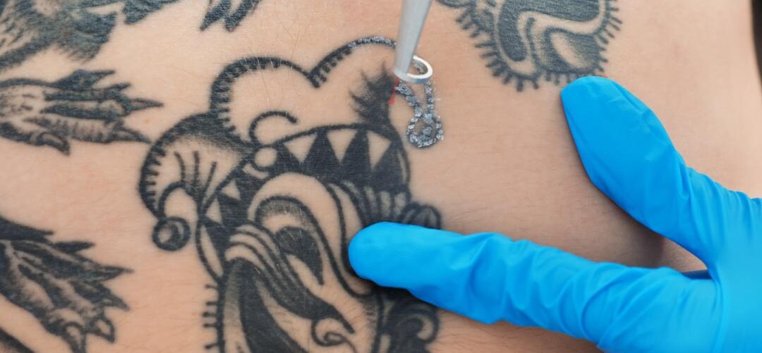
Safety Considerations for Practitioners
Safety considerations for practitioners performing laser tattoo removal are critical to ensure both patient and clinician well-being. Laser tattoo removal involves high-intensity light, tissue interactions, and potential chemical and immunologic responses, all of which carry inherent risks. A structured, evidence-based approach is essential to minimize complications and achieve predictable outcomes.
Protective measures for the clinician are paramount. Eye protection with wavelength-specific safety goggles is mandatory for both the operator and the patient, as inadvertent exposure to laser energy can cause permanent ocular injury. Proper room shielding, warning signage, and adherence to laser safety protocols reduce the risk of accidental exposure. Operators should also use appropriate personal protective equipment, including gloves and masks, to prevent contact with pigment-laden aerosols or debris released during treatment.
Patient safety begins with thorough pre-treatment assessment. This includes evaluating skin type, tattoo age, pigment colors, depth, and prior reactions. Patients should be screened for conditions that increase risk, such as photosensitivity disorders, autoimmune or dermatologic diseases, or a history of keloid formation. Informed consent is essential and should include discussion of potential complications, including paradoxical darkening, delayed hypersensitivity, infection, and scarring. Patients must understand that multiple sessions may be required and that results can vary depending on pigment type, depth, and composition.
Proper laser selection and parameter adjustment are key safety considerations. Practitioners should select wavelengths appropriate for the pigments being treated, and use conservative fluence and pulse durations to minimize thermal injury and inflammation. Test spots are recommended for tattoos of unknown composition or high-risk colors. Adequate cooling techniques, such as contact or cryogen-based systems, can reduce epidermal damage, discomfort, and the risk of blistering.
Infection control is essential. Even when inks are sterile, laser treatment can disrupt dermal tissue and introduce contaminants from the environment. Strict adherence to hand hygiene, surface disinfection, and proper handling of consumables reduces the risk of post-treatment infections. Practitioners must also educate patients on post-laser care, including wound hygiene, avoidance of sun exposure, and monitoring for signs of infection or adverse immune responses.
Monitoring and documentation are critical components of practitioner safety and quality control. Recording laser parameters, pigment characteristics, clinical observations, and patient responses helps identify patterns of adverse reactions and informs adjustments for subsequent sessions. This documentation is also important for medico-legal protection and ongoing clinical evaluation.
Practitioners must remain vigilant for systemic reactions, especially in cases involving large tattoos, multiple pigment types, or tattoos with known impurities. Rarely, laser-induced pigment fragmentation can result in transient systemic exposure, potentially provoking immune or hypersensitivity responses. Awareness of these risks allows early recognition and appropriate management.
The Role of Education and Evidence-Based Practice
 At Vancoderm Academy & College, education is grounded in scientific rigor, clinical competence, and evidence-based practice. Our programs in medical aesthetics and laser technologies emphasize not only procedural skills but also a deep understanding of underlying physiology, dermatologic principles, and the behavior of tattoo pigments in the skin.
In advanced laser tattoo removal training, (Clinical Practitioner Specialist Diploma in Medical Aesthetics) students learn to analyze pigment composition, particle size, dermal distribution, and the potential impact of impurities and carriers. The curriculum integrates knowledge of immune responses, chemical reactivity, paradoxical darkening, and regulatory considerations, preparing graduates to anticipate and manage complex clinical scenarios safely.
Hands-on clinical training is paired with theoretical instruction, ensuring students gain practical experience in laser selection, parameter adjustment, patient assessment, risk mitigation, and post-treatment care. Emphasis is placed on understanding the variability of tattoo inks, managing multicolor tattoos, and minimizing complications such as hypersensitivity, scarring, or infection.
Vancoderm Academy & College also prioritizes evidence-based decision-making. Students are trained to critically evaluate current research, integrate new findings into practice, and apply standardized protocols to achieve predictable, safe outcomes. Case studies, simulated treatment planning, and peer-reviewed literature form an integral part of the learning experience.
 Our graduates are equipped not only with technical proficiency but also with the knowledge to educate patients about risks, expected outcomes, and post-treatment care. By fostering both clinical competence and scientific understanding, Vancoderm Academy & College ensures that practitioners are prepared to deliver high-quality, safe, and professional laser tattoo removal services in a rapidly evolving field.
I would like to sincerely thank our readers for their continued support and engagement. Your interest in advancing knowledge in medical aesthetics and laser tattoo removal motivates us to share evidence-based insights and best practices. We also appreciate our followers on LinkedIn, Instagram, and Facebook, whose participation helps us build a community of informed professionals and learners. Your trust and enthusiasm inspire Vancoderm Academy & College to continue delivering high-quality educational content and clinical guidance.