
White Dots on Nail(Leukonychia)
 Faramarz Rafie MD / Vancoderm Academy and College [VDA] / Vancoderm Clinic [VDCMed]
It is not uncommon for patients to present with concerns regarding small white macules or linear streaks on the nail plate, often questioning whether these findings signify underlying systemic disease. In this week’s dermatology update from Vancoderm Academy & College, we examine this frequently encountered clinical observation—leukonychia—from an evidence-based, medical perspective. Our aim is to clarify the etiologic mechanisms behind these nail changes and outline when such findings warrant further clinical evaluation.
What Is Leukonychia?
Leukonychia is a clinical term referring to white discoloration of the nail plate resulting from abnormalities in nail matrix keratinization or alterations in the nail plate itself. The condition is categorized based on its clinical morphology and the distribution of the white opacity.
 1. Punctate Leukonychia
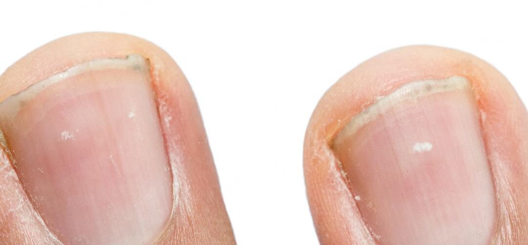
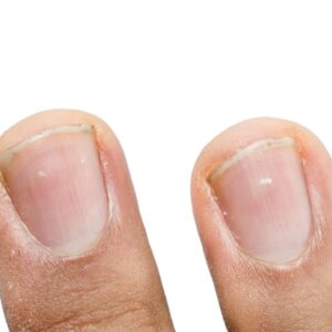
Punctate leukonychia is the most prevalent form and is characterized by small, discrete white spots within the nail plate.
-
These lesions typically arise from focal, transient trauma to the nail matrix, disrupting normal keratinocyte maturation.
-
Because the disrupted keratin becomes incorporated into the growing nail plate, the white spots migrate distally as the nail grows.
-
This variant is benign and does not indicate systemic disease.
 2. Striate (Longitudinal or Transverse) Leukonychia
Striate leukonychia presents as linear white bands, either horizontally (transverse leukonychia) or vertically (longitudinal leukonychia):
-
Transverse white bands, often referred to as Mees’ or Muehrcke-like lines depending on the underlying pathophysiology, may arise from episodic injury to the nail matrix, systemic illness, or medication exposure.
-
Longitudinal striae are less common and may be associated with inflammatory nail disorders or localized matrix pathology.
3. Partial or Total Leukonychia
In partial or total leukonychia, large sections—or the entirety—of the nail plate appear white:
-
Partial leukonychia involves segmental areas of opacity, often related to more extensive matrix disturbances.
-
Total leukonychia (leukonychia Totalis) signifies complete whitening of the nail plate and may be congenital or associated with systemic conditions, genetic syndromes, or profound matrix dysfunction. From a dermatologic standpoint, leukonychia is primarily a disorder of light reflection due to abnormal keratinization within the nail plate rather than a pigmentary problem. While punctate leukonychia is overwhelmingly benign and trauma-related, more extensive patterns may warrant further evaluation to exclude systemic or inherited causes.
Common Causes of White Dots on Nails
1. Minor Trauma to the Nail Matrix
The nail matrix, located proximally beneath the eponychium, is responsible for generating the nail plate through a tightly regulated process of keratinocyte proliferation and keratinization. Even subtle or repetitive mechanical insults to this region can transiently disrupt normal matrix function.
Common sources of microtrauma include:
-
Onychophagia (nail biting) and onychotillomania (nail picking): These behaviors exert intermittent pressure or shear forces on the proximal nail fold and matrix.
-
Aggressive or improper manicuring techniques: Excessive cuticle manipulation, mechanical trimming, or repeated use of abrasive instruments may irritate the matrix.
-
Sports-related impact injuries: Direct blows or repetitive micro-impacts to the distal digits can indirectly affect the matrix through transmitted force.
-
Tight or ill-fitting footwear (particularly relevant to toenails): Chronic compression leads to low-grade repetitive trauma to the matrix of the hallux and lesser toes.
When matrix keratinocytes are injured, parakeratotic or incompletely keratinized cells become incorporated into the forming nail plate. This results in focal areas of light scattering, clinically visible weeks later as punctate leukonychia as the nail grows distally. This trauma-induced leukonychia is benign, self-limiting, and non-indicative of systemic disease, with resolution occurring naturally as the nail plate advances.
2. Allergic or Irritant Reactions
Exposure to allergens or chemical irritants found in nail cosmetics can induce inflammatory changes in the proximal nail fold and matrix.
Common culprits include:
-
Acrylates and methacrylates in artificial nail systems
-
Toluene sulfonamide–formaldehyde resin in nail polishes
-
Solvents (e.g., acetone, ethyl acetate) in removers
-
Nail hardeners containing formaldehyde derivatives
In susceptible individuals, these agents may trigger allergic contact dermatitis (ACD) or irritant contact dermatitis (ICD) involving the periungual tissues. Inflammation of the matrix disrupts the normal keratinization gradient, leading to white macules or streaks in the nail plate (a form of acquired leukonychia). Unlike trauma-induced lesions, these changes may occur across multiple nails simultaneously if the allergen exposure is generalized.
3. Nutritional Misconceptions
Although widely believed by the public, simple white dots on the nails rarely indicate mineral or vitamin deficiency.
True nutrition-related nail pathology typically manifests differently:
-
Zinc deficiency: may cause Beau’s lines, onychorrhexis, or diffuse plate thinning
-
Iron deficiency (anemia): associated with koilonychia, not leukonychia
-
Calcium deficiency: does not manifest as leukonychia
-
Protein-energy malnutrition: can lead to brittle nails or slow growth, not punctate white spots
White dots are far more often caused by focal matrix trauma than micronutrient abnormalities. Thus, routine supplementation is not indicated unless corroborated by systemic signs or laboratory evidence.
4. Medication- or Illness-Related Matrix Disturbance
More extensive or patterned leukonychia may be linked to systemic factors that interrupt nail matrix metabolism. These typically present as transverse white bands rather than isolated dots.
Potential contributors include:
-
Systemic illnesses accompanied by high fever (e.g., severe infections)
-
Nephrotic syndrome or hepatic dysfunction (associated with Muehrcke lines)
-
Chemotherapeutic agents (Taxanes, Anthracyclines, Vincristine), Taxanes refers to a class of plant-derived chemotherapeutic agents, including paclitaxel and docetaxel, which are widely utilized in the treatment of breast, lung, prostate, and other solid malignancies. These agents function by disrupting microtubule dynamics, thereby inhibiting cell division and tumor proliferation.
-
Heavy metal exposure (e.g., arsenic, thallium—classically producing Mees’ lines)
These conditions lead to temporary arrest or alteration in nail matrix keratinization, producing white, often symmetric, lines across several nails. Unlike punctate leukonychia, these patterns may hold diagnostic value and warrant evaluation of the patient’s systemic history.
5. Fungal Infections (Uncommon Cause of White Dots)
Although fungal nail infections (onychomycosis) are common, they rarely present as isolated white dots. The exception is superficial white onychomycosis (SWO), caused by organisms such as Trichophyton mentagrophytes.
Features include:
-
Diffuse chalky white patches on the surface of the nail plate
-
Surface friability when gently scraped
-
Possible spread with coexisting tinea pedis
Because SWO represents fungal colonization of the superficial nail plate, it does not produce discrete matrix-derived punctate leukonychia. Diagnosis is supported by KOH preparation, fungal culture, or PAS stain, and treatment may involve topical antifungals or oral therapy in refractory cases.
6. When Leukonychia Warrants Clinical Evaluation
While most cases of punctate leukonychia are benign, evaluation is recommended if the following are observed:
-
Generalized leukonychia affecting several or all nails
-
Accompanying nail plate abnormalities such as thickening, splitting, or surface distortion
-
Rapid onset following systemic symptoms or new medications
-
Longitudinal leukonychia (which may indicate matrix neoplasia or inflammatory pathology)
-
Lack of growth or persistence despite nail plate advancement
In such cases, a comprehensive dermatologic assessment—including review of systemic health, medication exposure, occupational hazards, and potential genetic conditions—may be indicated.
When Should You Seek Medical Attention?
Although punctate leukonychia is typically benign and self-limited, certain patterns warrant further clinical evaluation. Patients should seek assessment by a dermatologist or certified nail specialist at Vancoderm Academy & College if any of the following features are present:
-
Diffuse or generalized white discoloration involving multiple or all nails, which may suggest systemic involvement or a hereditary nail disorder.
-
Concurrent nail plate abnormalities such as onycholysis, onychorrhexis, splitting, thickening, or periungual inflammation, which may indicate infection, inflammatory nail disease, or structural matrix pathology.
-
Rapid progression or sudden onset of nail changes without clear history of trauma, raising suspicion for systemic triggers, toxic exposures, or drug-related effects.
-
Past or current systemic illness, including renal, hepatic, hematologic, or autoimmune conditions, which can manifest with characteristic nail findings such as Muehrcke’s or Mees’ lines.
Persistent, unusual, or widespread leukonychia may serve as a clinical marker of underlying systemic dysfunction, and a targeted dermatologic evaluation can help determine whether further laboratory or diagnostic workup is indicated.
How to Care for and Prevent White Dots on Nails
Preventive nail care focuses on minimizing trauma to the nail matrix and reducing exposure to irritants and allergens. Recommended strategies include:
-
Avoid aggressive grooming practices, such as excessive cuticle manipulation, forceful trimming, or picking at the proximal nail fold, all of which can disrupt matrix integrity.
-
Use protective gloves during household cleaning, wet work, or manual labor to reduce mechanical and chemical insults to the nails and periungual skin.
-
Select hypoallergenic nail products, including polishes and removers free of common sensitizers such as acrylates and formaldehyde resins, particularly for individuals with a history of contact dermatitis.
-
Provide periodic rest from cosmetic nail enhancements, including gel or acrylic systems, to allow natural recovery of the nail plate and matrix.
-
Maintain nail hydration with regular application of cuticle oils or emollients, which support barrier function and improve nail plate flexibility.
While nails can serve as valuable indicators of systemic and dermatologic health, most isolated white spots are benign. Thoughtful nail care combined with awareness of concerning features can help distinguish harmless changes from those requiring professional evaluation.
 About Vancoderm Academy & College
As a Designated Learning Institution (DLI) in Canada, Vancoderm Academy & College is recognized as a leading provider of advanced medical aesthetics education. Our flagship Clinical Practitioner Specialist Diploma in Medical Aesthetics offers a comprehensive, evidence-based curriculum that integrates clinical dermatology, cosmetic science, and hands-on procedural training. This unique and globally respected diploma program prepares graduates with the theoretical depth and practical competency required to excel in today’s rapidly evolving medical aesthetics field.
We extend our sincere appreciation to all our readers, students, and professional followers for your continuous support. We invite you to stay connected with Vancoderm Academy & College by following us on Instagram, LinkedIn, Facebook, and all major social platforms for ongoing updates, educational content, and program announcements.
Wishing you and your families the very best during the upcoming Christmas season, and we look forward to continuing our commitment to excellence in dermatology and medical aesthetics education in the new year.
Thank you.