

Every Monday, we at #Vancoderm_Academy publish an insightful #blog aimed at enhancing our students’ knowledge and skills on specific #medical_aesthetic cases. This initiative is an integral part of our students’ learning journey during their studies in the #Clinical_Practitioner_Specialist_Diploma_in_Medical_Aesthetics™
While Dermatochalasis and Blepharoptosis (Ptosis) are distinct conditions, they frequently coexist, especially in older adults. Dermatochalasis can mask underlying ptosis, and ptosis may become more apparent after dermatochalasis is corrected (e.g., post-blepharoplasty). Therefore, a careful preoperative evaluation is essential.
In dermatochalasis cases, Botox should be approached with functional anatomy in mind, not just cosmetic goals. The goal is to avoid disrupting compensatory mechanisms that are helping the patient maintain their visual field.
To begin, I will provide an overview of dermatochalasis and blepharoptosis.
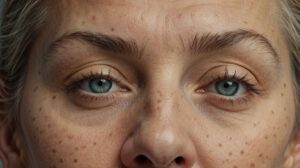
Dermatochalasis is a common age-related or hereditary condition characterized by redundant, lax skin of the upper and/or lower eyelids, often accompanied by orbital fat prolapse due to weakening of the orbital septum. The pathophysiology involves attenuation of dermal connective tissues, particularly elastin and collagen, leading to excessive skin redundancy. Clinically, dermatochalasis presents as draping upper eyelid skin with a normal eyelid margin position (normal MRD1). It may result in pseudoptosis or functional visual field obstruction, most commonly in the superior-temporal quadrant. Management is primarily surgical, with upper eyelid blepharoplasty indicated for both functional and cosmetic correction, sometimes including resection of herniated fat.
When considering botulinum toxin injection (Botox) in patients with dermatochalasis, several precautions are critical. Many of these patients rely on frontalis muscle activity to compensate for upper lid heaviness; Botox-induced relaxation of this muscle can unmask or worsen eyelid ptosis, impairing vision.
Injections should be placed high on the forehead and away from the orbital rim to preserve brow elevation and avoid unintended weakening of periorbital muscles. Additional risks include brow descent, which may exacerbate the hooded appearance of the eyelids.
Careful patient selection and informed consent are essential, as Botox may not be appropriate in all cases—particularly those with borderline ptosis or significant functional impairment—where surgical intervention may be more beneficial.
Upper Eyelid Ptosis (Blepharoptosis)
is defined as a pathological lowering of the upper eyelid margin, typically diagnosed by a reduced margin reflex distance 1 (MRD1), often less than 2 mm. It results from impaired function or structural disruption of the levator palpebrae superioris muscle or its innervation. The most common etiology in adults is aponeurotic (involutional) ptosis, caused by age-related stretching or disinsertion of the levator aponeurosis. Other causes include neurogenic (e.g., third nerve palsy, Horner’s syndrome), myogenic (e.g., myasthenia gravis, congenital myopathies), mechanical (e.g., tumors, edema), and traumatic factors.
Clinically, ptosis presents with a low-positioned upper eyelid, often accompanied by compensatory frontalis muscle recruitment, resulting in eyebrow elevation. In neurogenic cases, signs such as anisocoria or ophthalmoplegia may be present.
Management is etiology-specific:
levator advancement is preferred for aponeurotic ptosis, while levator resection or frontalis suspension is used in congenital cases. Neurogenic and myogenic ptosis require treatment of the underlying disorder, with surgery considered in stable or non-responsive cases. Accurate diagnosis and classification are essential for selecting the appropriate therapeutic approach.
Precautionary Evaluation in Patients with Dermatochalasis to Detect or Prevent Missed Ptosis
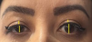
In patients with dermatochalasis, a thorough preoperative eyelid assessment is essential to distinguish true ptosis from pseudoptosis and to avoid postoperative surprises. Begin with measurement of MRD1 (normal: ~4–5 mm; <2 mm suggests ptosis) and levator function (normal: >12 mm; poor: <4 mm, often requiring frontalis suspension).
Differentiate pseudoptosis by manually lifting redundant upper eyelid skin—if the lid margin normalizes, it’s dermatochalasis alone; if it remains low, true ptosis is present. Assess brow position and look for compensatory frontalis overaction, which may mask underlying ptosis; instruct the patient to relax the brow for accurate evaluation. Visual field testing (e.g., Humphrey or Gold
mann perimetry) is crucial for documenting functional visual obstruction, particularly when surgery is medically indicated.
Additional considerations include fatigue or diurnal variability, which may indicate myasthenia gravis—a fatigue test (sustained upgaze for 1–2 minutes) may help unmask fluctuating ptosis.
Apply Hering’s Law when ptosis is bilateral, as unilateral correction can unmask contralateral ptosis, often necessitating bilateral intervention. Perform preoperative photography to document eyelid position and counsel patients that ptosis may become more apparent after blepharoplasty and may require a second-stage procedure. Evaluate for neurogenic or myogenic etiologies—refer to neurology or ophthalmology if conditions such as myasthenia gravis, Horner’s syndrome, or cranial nerve III palsy are suspected. Before any Botox injection or eyelid surgery, repeat measurements of MRD1 and levator function, evaluate brow dynamics, assess for compensatory mechanisms, and document all findings thoroughly.
Technical Considerations and Precautions for Botox Use in Dermatochalasis
Injecting Botox in patients with dermatochalasis requires careful planning and technique to avoid functional and cosmetic complications. Key precautions include assessing baseline measurements such as MRD1, levator function, and brow position before treatment.
It’s critical to avoid over-relaxing the frontalis muscle, as many patients use it to compensate for drooping eyelids. Injections should be placed high on the forehead (at least 2 cm above the brow) to preserve brow elevation, and lateral brow injections should be avoided to prevent additional brow descent.
Infraorbital orbicularis injections can weaken lower lid support and should also be avoided. A lower total dose (around 20–30 units) is recommended, particularly for new or borderline patients.
Individual asymmetry should be accounted for, as dermatochalasis can vary between the two sides. Patients should be informed of potential risks, including temporary brow or eyelid droop, especially in those with borderline ptosis. A follow-up evaluation after two weeks is essential to assess results and address any complications.
For a typical patient aged 45–55 with mild dermatochalasis, a conservative injection plan might include 6–10 units in the forehead (placed high), 10–12 units in the glabella, and 4–6 units per side in the crow’s feet area.
With cautious dosing and precise placement, Botox can still provide cosmetic benefits without exacerbating eyelid issues.
Faramarz Rafie M.D.
Vancoderm Academy