
 Vancodermadmin
Photodermatitis in Medical Aesthetics: Pathophysiology, Clinical Recognition, Treatment Risks, and Prevention
 Faramarz Rafie MD / Vancoderm Academy and College [VDA] / Vancoderm Clinic [VDCmed]
Vancoderm Academy and College of Aesthetic Medicine and Wellness | Weekly Educational Blog
Introduction
Photosensitivity is an important consideration in dermatology and medical aesthetics because the interaction between light, skin chromophores, medications, topical chemicals, and individual biological susceptibility can produce a wide range of cutaneous reactions.
For medical aesthetic professionals, this subject is particularly relevant when assessing clients for laser hair reduction, intense pulsed light (IPL), pigment-targeting procedures, chemical peels, and skin-resurfacing treatments. A client may present with redness, itching, burning, or persistent pigmentation that appears to be a routine skin concern but may reflect an underlying photosensitive disorder or a reaction to a photosensitizing agent.
Understanding these conditions requires more than recognizing visible inflammation. It requires knowledge of skin physiology, the mechanisms of phototoxicity and photoallergy, clinical distribution patterns, medication-related risks, differential diagnosis, and the limitations of aesthetic assessment.
The central principle is that unexplained inflammation or suspected photosensitivity must be assessed before elective medical aesthetic treatment. Recognizing a possible disorder and referring appropriately are essential professional competencies.
1. Definition and Medical Classification
What is photodermatitis?
Photodermatitis is an inflammatory skin reaction induced or aggravated by exposure to light, particularly ultraviolet (UV) radiation. The term is often used for eczematous or dermatitis-like reactions associated with photosensitivity.
The broader term photodermatosis describes skin disorders that are triggered or exacerbated by light. These include inflammatory eruptions, phototoxic reactions, photoallergic dermatitis, and other conditions that may present with blistering, urticaria, or abnormal pigmentation.
Photosensitivity itself describes an increased or abnormal response to light; it does not necessarily mean that the individual has photodermatitis.
Major categories of photosensitive disorders
1. Exogenous photodermatoses
These occur when an external substance interacts with light to produce a skin reaction. Examples include drug-induced phototoxicity, drug-induced photoallergy, and photocontact dermatitis.
2. Primary or idiopathic photodermatoses
These include disorders such as polymorphic light eruption and solar urticaria, in which abnormal responses to light occur without a single identifiable external photosensitizer explaining every case.
3. Photoaggravated dermatoses
Certain pre-existing conditions, including cutaneous lupus erythematosus and some cases of atopic dermatitis or rosacea, may worsen with light exposure.
4. Metabolic and inherited photosensitivity disorders
Porphyrias and certain rare genetic disorders can produce photosensitivity through mechanisms involving accumulated photoreactive substances or impaired cellular responses to environmental damage.
These categories may overlap clinically. Accurate diagnosis depends on the history, examination, and, when necessary, specialist investigations.
2. Pathophysiology: How Does Light Produce Skin Inflammation?
Photodermatitis is best understood by examining the interaction between incident radiation and molecules within or on the skin.
A. The role of ultraviolet radiation
Ultraviolet radiation is divided into three wavelength ranges:
- UVA: 315–400 nm. UVA penetrates into the dermis and contributes to oxidative stress, photoaging, and certain medication-induced photosensitive reactions. It can pass through ordinary untreated window glass to a significant extent.
- UVB: 280–315 nm. UVB is absorbed predominantly within the epidermis and is strongly associated with erythema, sunburn, and direct DNA damage.
- UVC: 100–280 nm. Solar UVC is absorbed by the atmosphere and does not ordinarily reach the Earth’s surface in significant amounts. Artificial UVC sources can nevertheless cause tissue injury.
Visible light can also trigger certain photosensitive disorders. The wavelengths responsible depend on the condition and the relevant photosensitizing molecule.
B. Phototoxicity: direct photochemical injury
Phototoxic reactions occur when a chemical present in the skin absorbs photons of an appropriate wavelength. The molecule enters an excited state and may transfer energy to oxygen or participate in electron-transfer reactions.
These processes can generate reactive oxygen species and free radicals, resulting in oxidative stress, membrane damage, inflammation, and cellular injury.
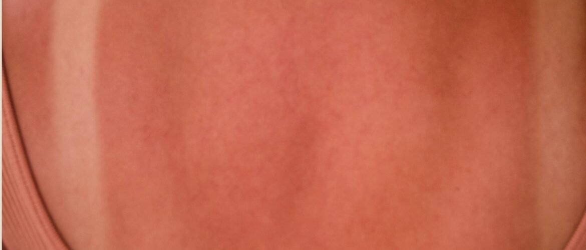
The clinical reaction may resemble an exaggerated sunburn, with erythema, edema, burning, tenderness, and sometimes blistering.
Phototoxicity is generally non-immunologic. It can occur on first exposure when sufficient quantities of the photosensitizing substance and an appropriate dose of light are present.
C. Photoallergy: delayed immune-mediated inflammation
Photoallergic dermatitis follows a different mechanism. Light alters a chemical or promotes its binding to skin proteins, producing a photoallergen that can initiate an immune response.
After sensitization, re-exposure may activate a delayed, T-cell-mediated type IV hypersensitivity reaction.
Clinical findings commonly include itching, eczematous redness, papules, vesicles, scaling, and sometimes weeping or crusting. The eruption may extend beyond the skin directly exposed to light.
Unlike phototoxicity, photoallergy requires sensitization and is not simply determined by the amount of light exposure.
D. Why this distinction matters
Although phototoxicity and photoallergy can both produce inflammation, their mechanisms, timing, distribution, and preventive approaches differ.
Recognizing the distinction can help guide clinical investigation, but appearance alone is not sufficient to establish a definitive diagnosis.
3. Etiology and Contributing Factors
A. Drug-induced photosensitivity
A range of systemic and topical medications can increase sensitivity to UV radiation or, less commonly, visible light.
Examples include certain:
- Tetracycline antibiotics, including doxycycline.
- Fluoroquinolone antibiotics.
- Non-steroidal anti-inflammatory drugs (NSAIDs).
- Thiazide and related diuretics.
- Antifungal medications, including selected azole drugs.
- Antiarrhythmic medications such as amiodarone.
- Retinoids and selected anticancer therapies.
The potential for photosensitivity differs substantially between medications. It also depends on the dose, duration, route of administration, concurrent products, light spectrum, and individual susceptibility.
A medication list should therefore be reviewed systematically rather than treated as a simple yes-or-no screening question.
Clinical safety point: Clients should not discontinue, reduce, or alter prescribed medication independently. If a potential medication-related risk is identified, the client should consult the prescribing healthcare professional and follow the applicable treatment-device guidance.
B. Topical products and photocontact dermatitis
Photocontact dermatitis occurs when a substance on the skin interacts with light to produce inflammation.
Potential sources include selected fragrances, topical medications, some sunscreen ingredients in susceptible individuals, disinfectants, and plant-derived chemicals.
A well-known example is phytophotodermatitis caused by contact with furocoumarin-containing plants, including lime, followed by exposure to UVA. This can produce sharply defined erythema, linear or irregular blistering, and prolonged post-inflammatory hyperpigmentation.
Not every cosmetic reaction is photo-induced. Irritant dermatitis, ordinary allergic contact dermatitis, and direct chemical injury must also be considered.
C. Underlying medical conditions
Photosensitivity may be associated with autoimmune diseases, metabolic abnormalities, inherited disorders, or chronic inflammatory skin disease.
A client who reports recurrent unexplained eruptions after minimal sunlight exposure, persistent photosensitivity, painful skin reactions, or unusual blistering should receive appropriate medical evaluation before elective procedures are considered.
D. Environmental and behavioural factors
Relevant factors include prolonged sunlight exposure, tanning, insufficient photoprotection, repeated exposure to a known trigger, and failure to follow post-procedure instructions.
The risk is not determined by sunlight alone. The interaction between the exposure, the individual’s biology, and the procedure or substance involved is what matters.
4. Clinical Signs and Symptoms
Clinical manifestations depend on the underlying disorder, radiation spectrum, intensity and duration of exposure, and the individual’s skin response.
Common findings
- Erythema, or visible redness.
- Edema and localized swelling.
- Burning, stinging, tenderness, or pain.
- Pruritus, or itching.
- Papules and eczematous plaques.
- Vesicles or bullae in more severe reactions.
- Scaling, dryness, or crusting.
- Post-inflammatory hyperpigmentation or, less commonly, hypopigmentation.
- Persistent skin thickening or lichenification in chronic inflammatory conditions.
Photodistribution
Photosensitive eruptions often affect exposed areas such as the face, V-shaped upper chest, neck, dorsal hands, forearms, and lower legs.
Relative sparing of shaded areas, skin beneath a watch strap, or areas protected by clothing can provide a useful clinical clue. However, these patterns are not universal.
Photoallergic dermatitis may spread beyond directly exposed areas, and some photodermatoses have distinctive distributions that do not follow a simple sun-exposed pattern.
Timing of onset
| Feature | Phototoxic reaction | Photoallergic reaction |
|---|---|---|
| Mechanism | Direct photochemical injury | Delayed immune-mediated hypersensitivity |
| Onset | Often minutes to hours | Commonly 24–72 hours after re-exposure |
| Sensitization required | No | Yes |
| Typical symptoms | Burning, pain, sunburn-like redness | Itching and eczematous inflammation |
| Distribution | Usually confined to exposed areas | Exposed areas, sometimes extending beyond them |
| Pigmentary aftermath | Possible and sometimes prominent | Possible, particularly after persistent inflammation |
These are typical patterns, not absolute rules. Atypical presentations and other diagnoses are possible.
5. Differential Diagnosis: What Else Can Look Similar?
Photodermatitis should not be diagnosed from redness or pigmentation alone. Several conditions can produce overlapping findings.
Sunburn: Direct UV-induced skin injury that commonly produces erythema and tenderness. It is not synonymous with photoallergic dermatitis.
Polymorphic light eruption: An inflammatory eruption that commonly develops hours to days after sun exposure and may present with itchy papules, plaques, or small blisters.
Solar urticaria: An uncommon condition characterized by rapid development of wheals after exposure to the relevant light wavelengths.
Allergic contact dermatitis: An eczematous reaction to a contact allergen that does not require light exposure.
Irritant contact dermatitis: Direct barrier injury caused by a substance such as an irritant cosmetic or chemical.
Rosacea or other photoaggravated dermatoses: Pre-existing conditions that may worsen with sunlight, heat, or other environmental triggers.
Cutaneous lupus erythematosus and dermatomyositis: Conditions that may produce photosensitive eruptions and require medical assessment.
Porphyria-related photosensitivity: Certain porphyrias can cause pain, blistering, fragility, or other characteristic reactions to light.
Persistent, recurrent, blistering, or unexplained photosensitive eruptions require appropriate medical evaluation. Depending on the presentation, a dermatologist may consider phototesting, photopatch testing, laboratory investigations, or skin biopsy.
6-Diagnosis of Photodermatitis: Clinical Assessment and Confirmatory Investigations
Accurate diagnosis of photodermatitis requires a systematic clinical assessment to establish whether a skin reaction is related to light exposure and to identify the underlying mechanism. Although the appearance and distribution of the eruption can provide important clues, no single clinical sign or laboratory test confirms every form of photodermatitis.
Diagnosis should be performed by an appropriately qualified healthcare professional, often a dermatologist when the presentation is persistent, recurrent, severe, or unclear.
1. Clinical History and Skin Examination
The diagnostic process begins with a detailed medical history and examination of the affected skin.
The healthcare professional will assess:
- Exposure history: The timing and duration of sunlight or artificial light exposure before symptom onset.
- Medication history: Prescription medications, over-the-counter drugs, supplements, and topical treatments that may cause photosensitivity.
- Product exposure: Recent use of cosmetics, fragrances, sunscreens, topical medications, or plant-derived substances that may cause photocontact reactions.
- Clinical presentation: The presence of erythema, edema, itching, burning, papules, vesicles, blisters, scaling, or pigmentary changes.
- Distribution: Whether the eruption predominantly affects light-exposed areas, with relative sparing of skin protected by clothing or other objects.
- Previous episodes: Recurrence, seasonal patterns, previous reactions to aesthetic procedures, and relevant medical or dermatological conditions.
The timing of the reaction can help distinguish possible mechanisms. Phototoxic reactions often develop within minutes to hours of exposure, whereas photoallergic reactions commonly appear after a delay of approximately 24–72 hours following re-exposure in a sensitized individual. These patterns are helpful clinical clues but are not definitive diagnostic criteria.
2. Phototesting
Phototesting is a specialist investigation used to evaluate abnormal skin responses to specific wavelengths of light.
During the procedure, small areas of skin are exposed to controlled doses of ultraviolet A (UVA), ultraviolet B (UVB), and, when appropriate, visible light. The skin response is assessed at specified intervals.
Depending on the suspected condition, phototesting may help to:
- Determine whether the individual has an abnormal response to light.
- Identify the wavelengths associated with the reaction.
- Measure the minimal erythema dose (MED), the lowest UV dose that produces a defined erythematous response under specified testing conditions.
- Investigate suspected photodermatoses, including selected cases of chronic actinic dermatitis or polymorphic light eruption.
An abnormal phototest result must be interpreted in the context of the patient’s history and examination. Phototesting does not independently establish every type of photodermatitis.
3. Photopatch Testing
Photopatch testing is particularly useful when photoallergic contact dermatitis is suspected.
In this procedure, suspected contact allergens are applied to the skin, usually on the back, in two corresponding sets. After an initial application period, one set is exposed to a controlled dose of UVA, while the other remains unirradiated as a comparison.
The sites are subsequently examined for allergic reactions.
A positive reaction at the irradiated site, with a negative or substantially weaker reaction at the unirradiated site, may support a photoallergic response to the tested substance. Reactions at both sites may indicate ordinary allergic contact dermatitis, although interpretation depends on the complete test pattern.
Photopatch testing may be considered when a reaction is associated with selected sunscreen ingredients, topical medications, fragrances, or other suspected photocontact allergens. Results must be interpreted by a clinician with appropriate expertise.
4. Skin Biopsy and Histopathological Examination
A skin biopsy may be indicated when the clinical presentation is atypical or when other inflammatory, autoimmune, or blistering disorders must be excluded.
A small skin specimen is examined microscopically to assess the pattern of inflammation and other tissue changes.
Histopathological findings may help distinguish among possible diagnoses, but they are not always specific to photodermatitis. A biopsy must therefore be interpreted alongside the clinical findings and, when appropriate, phototesting or laboratory investigations.
5. Targeted Laboratory Investigations
Laboratory tests are selected according to the suspected underlying cause rather than ordered routinely for every patient.
Examples include:
- Autoimmune investigations: Tests such as antinuclear antibodies (ANA) and anti-Ro/SSA antibodies may be considered when cutaneous lupus erythematosus or another autoimmune disorder is suspected.
- Porphyrin studies: Blood, urine, or stool porphyrin testing may be indicated when a porphyria-related photosensitivity disorder is suspected.
- Additional investigations: Other blood tests or specialist assessments may be required based on the patient’s symptoms, examination, and medical history.
These tests investigate specific underlying conditions; they do not serve as universal confirmatory tests for photodermatitis.
6. Differential Diagnosis
Before confirming photodermatitis, the healthcare professional may need to distinguish it from other conditions with similar clinical features, including:
- Ordinary sunburn.
- Irritant or allergic contact dermatitis.
- Polymorphic light eruption.
- Solar urticaria.
- Photoaggravated rosacea or other inflammatory skin diseases.
- Cutaneous lupus erythematosus.
- Porphyria-associated photosensitivity.
- Other medication-related skin eruptions.
The differential diagnosis is important because the appropriate treatment, prognosis, and preventive measures vary considerably among these conditions.
7. What Should Medical Aesthetics Professionals Do?
Medical aestheticians play an important role in recognizing potential photosensitivity during pre-treatment consultations. However, screening is not equivalent to making a medical diagnosis.
If a client presents with unexplained redness, itching, blistering, recurrent reactions to sunlight, or unusual pigmentation associated with a suspected photosensitive eruption, the practitioner should:
- Document the client’s reported symptoms, relevant exposures, medication history, and observed skin findings.
- Defer elective medical aesthetic procedures when active inflammation or a potentially significant photosensitivity risk is present.
- Recommend assessment by an appropriate healthcare professional.
- Follow the clinic’s referral, documentation, and adverse-event procedures.
- Proceed with treatment only when suitability has been appropriately established and applicable device instructions and clinical protocols are satisfied.
Clients should not be advised to discontinue prescribed medications without consulting their prescribing healthcare professional.
Clinical takeaway: The confirmation of photodermatitis depends on integrating the clinical history, skin examination, and appropriate investigations. Phototesting and photopatch testing can provide valuable evidence in selected cases, while biopsy and laboratory studies may help identify or exclude specific underlying disorders.
For medical aesthetics professionals, the essential responsibility is to recognize possible photosensitivity, avoid treating unsuitable skin, and facilitate appropriate medical assessment before proceeding.
7. Risks in Medical Aesthetic Procedures
Photodermatitis is relevant to aesthetic practice because procedures may introduce additional optical energy, inflammation, or disruption of the skin barrier.
However, the level of risk differs between procedures. An active photosensitive eruption, a history of drug-induced photosensitivity, and a previous episode of sunburn are not interchangeable clinical findings.
A. Laser hair reduction and IPL
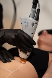
Laser and IPL devices deliver optical energy that is absorbed by target chromophores, producing intended thermal or photochemical effects.
A client with active dermatitis, a relevant photosensitizing medication, or an unexplained photosensitive reaction may be at increased risk of excessive inflammation, burns, blistering, pigmentary changes, or delayed recovery.
The specific risk depends on the device’s wavelength or spectrum, pulse duration, fluence, spot size, cooling system, treatment area, and client-specific factors.
A medication-associated photosensitivity reaction does not automatically establish a permanent contraindication to every laser or IPL procedure. Nevertheless, active symptoms or a suspected clinically significant interaction require assessment and may warrant postponement and referral.
B. Pigment-targeting lasers and tattoo removal
Pigment-targeting procedures deliver energy to selected chromophores, including melanin or tattoo pigment.
Unrecognized inflammation, recent tanning, altered skin reactivity, or a history of pigmentary complications may affect treatment suitability and increase the risk of post-inflammatory dyspigmentation.
Tattoo removal also has its own procedure-specific risks. Photodermatitis should be assessed as one element of the client’s overall clinical and treatment history.
C. Chemical peels
Chemical peels produce controlled chemical injury at a depth determined by the formulation, concentration, pH, application technique, and other factors.
Inflammation and temporary barrier impairment can increase the likelihood of irritation and pigmentary complications in susceptible clients. Active dermatitis or an unexplained photosensitive eruption should be assessed before proceeding.
Aftercare must address barrier recovery and photoprotection, with instructions appropriate to the depth and type of peel.
D. Microneedling and resurfacing
Microneedling creates controlled microchannels in the skin. Other resurfacing procedures may also induce inflammation or temporarily compromise the barrier.
Treating actively inflamed, blistered, or unexplained lesions may aggravate the condition or complicate healing. The underlying cause should be assessed before treatment.
E. Radiofrequency and other energy-based procedures
Radiofrequency procedures primarily produce tissue heating rather than relying on UV radiation. Therefore, they should not automatically be classified as light-induced procedures.
Nevertheless, active dermatitis, impaired barrier function, and an unexplained inflammatory condition may still affect treatment suitability. Device-specific contraindications and the client’s overall clinical status must guide the decision.
Clinical principle: A procedure’s risk should be evaluated according to its mechanism, the client’s condition, and the manufacturer’s instructions—not simply because the procedure is described as an aesthetic treatment.
8. Clinical Assessment Before Treatment
A structured pre-treatment assessment is an essential part of safe aesthetic practice.
Step 1: Obtain a relevant history
Ask about:
- Previous photosensitive eruptions or unusual reactions to sunlight.
- Current prescription and non-prescription medications.
- Topical medications, cosmetics, fragrances, and recent skincare changes.
- Recent sun exposure or tanning.
- Previous laser, IPL, peel, or resurfacing complications.
- Known dermatological conditions and relevant medical diagnoses.
- The onset, duration, recurrence, and progression of current symptoms.
Step 2: Examine the treatment area
Observe for erythema, edema, scaling, vesicles, bullae, crusting, skin fragility, or unexplained pigmentary changes.
Assess the distribution and whether it corresponds to light exposure, product application, clothing patterns, or the proposed treatment area.
Photographic documentation may be useful when appropriate and consented to, but it does not replace medical diagnosis.
Step 3: Evaluate suitability
If active inflammation, suspected photodermatitis, unexplained blistering, or a potentially significant medication-related risk is identified, do not proceed automatically.
Follow the clinic’s escalation protocol, relevant device instructions, and scope-of-practice requirements. Obtain appropriate medical assessment when indicated.
Step 4: Document and communicate
Record relevant history, observations, the decision to proceed or defer, any referral recommendation, and the advice given to the client.
Clear documentation supports continuity of care and responsible professional practice.
9. Prevention and Photoprotection
Prevention should address both the underlying photosensitivity and the risks associated with the planned procedure.
Before treatment
Medication review: Identify potential photosensitizing medications and consult the prescribing healthcare professional when the risk or appropriate waiting period is uncertain.
Skin assessment: Defer elective treatment when the skin has active, unexplained inflammation or suspected photodermatitis.
Sun-exposure history: Ask about recent tanning and sun exposure, and follow the specific device or procedure guidance.
Product review: Identify recently introduced topical products or potential photocontact triggers.
Informed consent: Explain relevant risks, expected recovery, aftercare requirements, and the importance of reporting unexpected reactions.
Device-specific protocols: Use the prescribed contraindication screening, parameter selection, cooling, protective eyewear, and treatment procedures for the particular device.
After treatment
Broad-spectrum protection: Recommend a broad-spectrum sunscreen with SPF 30 or higher, appropriate for the client and the procedure, applied according to the label and reapplied during outdoor exposure.
Physical photoprotection: Encourage shade, a wide-brimmed hat, and protective clothing. UVA can penetrate ordinary untreated window glass, so exposure through windows may also matter in selected photosensitive conditions.
Barrier care: Follow the procedure-specific recovery protocol and avoid introducing irritating products before the skin is ready.
Follow-up: Provide clear instructions about expected recovery and when the client should seek medical assessment.
Record keeping: Document the treatment parameters, client response, and any unexpected adverse event.
No single sunscreen or waiting period eliminates all photosensitivity risk. Prevention must be tailored to the underlying condition, relevant wavelength, exposure, and procedure.
10. Recognition of Adverse Reactions and Referral
Medical aesthetic professionals should be able to recognize a possible complication without exceeding their scope of practice.
Prompt medical evaluation is warranted for severe or worsening pain, extensive blistering, marked swelling, spreading inflammation, suspected infection, or an unexplained persistent eruption.
Emergency assistance is required for symptoms suggesting a severe systemic allergic reaction, such as difficulty breathing, collapse, or swelling of the tongue or throat.
When a reaction occurs after an aesthetic procedure:
- Stop the procedure or further exposure.
- Follow the clinic’s adverse-event and emergency protocols.
- Protect the affected area and avoid additional irritating treatments.
- Arrange medical assessment according to the severity and clinical findings.
- Document the timeline, exposure, device parameters, products used, and subsequent course.
- Review the event to identify preventive measures for future practice.
Treatment of confirmed photodermatitis depends on its cause and severity. Management may involve avoidance of the trigger, photoprotection, skin-barrier care, and clinician-directed medication. Aesthetic practitioners should not independently prescribe treatment for an undiagnosed skin reaction outside their authorized scope.
11. Clinical Case Example for Medical Aesthetic Diploma Students
A client books a laser hair reduction appointment and reports that, during the previous week, the backs of both hands developed redness and burning after outdoor exposure. The client recently started a new medication and has also noticed darker patches as the redness subsides.
On examination, the aesthetic professional observes inflammation with a distribution that appears more pronounced on exposed skin.
What should the practitioner consider?
- Could this represent a photosensitive drug reaction?
- Is the distribution consistent with a light-associated eruption?
- Is the medication known to have photosensitizing potential?
- Are there other possible causes, such as contact dermatitis or a different dermatological condition?
- Is it appropriate to perform an elective laser procedure before the cause is clarified?
Appropriate professional response: Defer the elective procedure, document the findings, advise the client to contact the prescribing healthcare professional, and recommend medical assessment as appropriate. Do not diagnose the reaction solely from its appearance or tell the client to stop the medication.
The purpose of this case is to demonstrate clinical reasoning, not to establish a diagnosis from limited information.
 12. Why This Knowledge Matters in Medical Aesthetics Education
Safe medical aesthetic practice requires the integration of skin science, client assessment, device knowledge, procedural technique, and recognition of adverse effects.
Understanding photodermatitis helps students appreciate why two clients receiving the same procedure may have different risk profiles. Medication history, current inflammation, recent sun exposure, underlying skin conditions, and individual pigmentary responses can all influence treatment planning.
At Vancoderm Academy and College of Aesthetic Medicine and Wellness, theoretical knowledge and practical skills are important components of medical aesthetics education. Students need to understand not only how a procedure is performed, but also why a treatment may need to be postponed, modified, or referred for further assessment.
Training in skin assessment, contraindication recognition, laser and light-based technologies, infection prevention, client communication, and adverse-effect awareness provides a foundation for responsible practice.
The objective is to develop professionals who approach medical aesthetic procedures with informed judgment, respect for scope of practice, and a commitment to client safety.
13. Vancoderm Academy Medical Aesthetics Diploma Program
Vancoderm Academy offers a Medical Aesthetics Diploma Program combining theoretical learning with practical training relevant to professional aesthetic practice.
Areas of study include skin assessment, cosmetic laser and light-based technologies, treatment protocols, contraindications, infection prevention, client consultation, and the recognition and prevention of potential complications.
The program emphasizes the importance of understanding skin physiology and treatment mechanisms alongside hands-on skills. Students learn why individual assessment, informed consent, appropriate documentation, and referral when indicated are integral to professional practice.
 Next scheduled intake: December 1, 2026.
Prospective students are encouraged to contact Vancoderm Academy for current information about program structure, admission requirements, schedules, and enrollment.
Vancoderm Academy and College of Aesthetic Medicine and Wellness
North Vancouver, British Columbia, Canada
Website: https://vancodermacademy.com/
Conclusion
Photodermatitis represents a clinically important group of light-associated inflammatory reactions with different mechanisms, presentations, and risk factors. Distinguishing phototoxic injury from photoallergic dermatitis and other photosensitive disorders is essential for appropriate investigation and prevention.
In medical aesthetics, a careful history, skin assessment, medication review, recognition of contraindications, and appropriate referral can help prevent avoidable complications. Sun protection and procedure-specific aftercare remain important, but they cannot replace the assessment of active or unexplained skin disease.
At Vancoderm Academy, the study of skin conditions such as photodermatitis reinforces a fundamental principle of professional education: technical proficiency must be supported by scientific understanding, clinical awareness, and responsible treatment decisions.